The optic nerve, which is known as the second cranial nerve, plays an essential role in helping us see. It carries the information collected by the retina and delivers it to the primary visual cortex in the brain, where our brain makes sense of what we are looking at.
Outside the brain (extracranial):
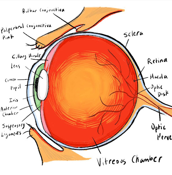
Inside the retina, special light-sensitive cells called photoreceptors turn light into tiny electrical signals. These signals are passed on to cells called bipolar cells, which release a chemical messenger called glutamate. This chemical activates the retinal ganglion cells, which create action potentials—tiny electrical impulses that are the language of our nervous system.
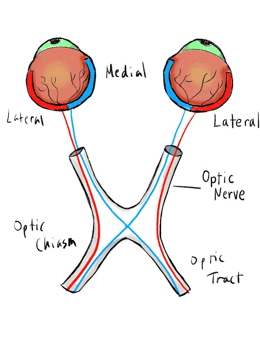
The long extensions (axons) of the retinal ganglion cells come together to form the optic nerve. This nerve leaves the protective bony eye socket through a small passage called the optic canal and then enters the skull’s middle cranial fossa, a space near the pituitary gland.
Inside the brain (intracranial):
Once inside the middle cranial fossa, the two optic nerves from each eye meet and merge at a structure called the optic chiasm. This crossing point sits right above a part of the sphenoid bone called the sella turcica. At the optic chiasm, the nerve fibers from the inner (medial) part of each retina cross over to the opposite side of the brain, while the fibers from the outer (lateral) part stay on the same side.
This arrangement means that information from the left side of what you see ends up traveling to the right side of your brain, and information from the right side goes to the left side.
After crossing (or not crossing) at the optic chiasm, these nerve pathways are called optic tracts. Each optic tract carries the visual information to a relay center deep in the brain called the lateral geniculate nucleus, or LGN, which is part of the thalamus. From the LGN, the signal is passed to the next set of neurons that send it along special pathways called optic radiations.
The lower parts of what we see are sent along the upper optic radiations, while the upper parts of our view travel along the lower optic radiations. Both paths lead to the primary visual cortex in the occipital lobe at the back of the brain—where the brain finally pieces everything together and creates the clear, detailed images we see every day.