Tuesday, March 25, 2025

The Life of Paul Farmer

I came across an article in the American Anthropologist Journal about medical anthropologist Paul Farmer. His life story is inspiring and pushes everyone to start thinking about how they can help others. Paul Farmer believed medicine had to start with people’s lives. Born in 1959, he grew up in unusual places like a converted school bus and a houseboat in Florida. He went to Duke University, where a medical anthropology class changed how he viewed medicine. Instead of studying science only in a lab, Farmer wanted to understand how history, politics, and culture shaped people’s health.

In the 1980s, he worked in rural clinics in Haiti, where malnutrition and preventable diseases were part of daily life. Rather than just observing, he built partnerships with local leaders such as Father Fritz Lafontant and the people of Cange. Together they founded Partners In Health (PIH), which became a global network proving that tuberculosis and HIV could be treated among the poor. Farmer called this approach a “preferential option for the poor.” It meant hiring and training local health workers who visited patients at home, brought medicine, and made sure basic needs like food and housing weren’t ignored.

Farmer was never afraid to call out injustice. At Harvard Medical School, he realized there were excess medical supplies in Boston hospitals while his patients in Haiti went without the basics. He called the deaths he witnessed “stupid deaths,” meaning deaths caused by diseases that were preventable and treatable. His books such as Pathologies of Power and Infections and Inequalities explained this as structural violence, showing how poverty and racism quietly but powerfully harm poor and vulnerable people.

Even as PIH expanded to countries like Peru and Rwanda, Farmer stayed close to his patients. In Boston, he treated the homeless, and in Africa, he helped fight the Ebola epidemic when many global health authorities had given up. Farmer believed that solidarity meant showing up everyday. He once said, “You just don’t leave.” That commitment inspired a generation of students and doctors to think differently about health as a human right.

Farmer was also known for his generosity and humility. He often gave away his possessions, even borrowing money just to hand it to someone else in need. Before an important meeting, he once fixed his only suit with a black marker. He cooked meals for family and took time to listen to everyone he met. After his lectures, he would stay for hours to answer questions from students and strangers.

When he died in Rwanda in 2022, Farmer was still doing what he had always loved: visiting patients, mentoring young doctors, and sharing jokes in the hospital wards. His gravestone reads, “Paul Farmer—Was of Use.” For the communities he served, that is more than true.

Tuesday, March 11, 2025

Evolutionary Impact of Social Isolation

One of the greatest mysteries in human evolution is how the Neanderthals went into extinction ~40 thousand years ago. This paper discusses the genome of a Neanderthal called “Thorin,” found in Grotte Mandrin in Mediterranean France. Thorin’s remains include a mandible and 31 teeth from between 52,900 and 48,050 years ago. These remains are linked to the PNII lithic industry, one of the final Neanderthal cultural phases in the region. Genomic analysis shows that Thorin belonged to a previously unknown Neanderthal lineage that diverged from other late European Neanderthals around 100,000 years ago and then remained genetically isolated for about 50,000 years. This isolation is unusual because other late Neanderthals were thought to be part of a single connected population.

Genetic analysis supports the isolation of this Neanderthal lineage. Thorin’s genome showed no signs of gene flow with other Neanderthals, including those living nearby. His mitochondrial DNA was most closely related to individuals from Gibraltar and Poland, and his Y chromosome also diverged early. Thorin also had high levels of homozygosity, with around 7% of his genome in long homozygous stretches, indicating recent inbreeding and a small group size. These findings suggest that his population had limited social contact with other Neanderthals. Interestingly, this isolated group also had a distinctive cultural identity, with stone tools that differed from the better-known Mousterian and Châtelperronian traditions in nearby areas.

The researchers conclude that Thorin's population likely represents one of the last Neanderthal groups in Europe, living in isolation until around 42,000 years ago. Their genetic and cultural separation may have made them more vulnerable to extinction. Unlike modern humans, who built wide social networks and exchanged genes across regions, these Neanderthal groups were fragmented and possibly avoided interbreeding—even with other Neanderthals. This paper adds more knowledge to the debate about Neanderthal extinction, suggesting that social and genetic isolation may have been just as important as external factors like climate or modern human expansion.

This is the paper I read: https://www.cell.com/cell-genomics/fulltext/S2666-979X(24)00177-0

Friday, February 28, 2025

The Optic Pathway

The optic nerve, which is known as the second cranial nerve, plays an essential role in helping us see. It carries the information collected by the retina and delivers it to the primary visual cortex in the brain, where our brain makes sense of what we are looking at.

Outside the brain (extracranial):
Inside the retina, special light-sensitive cells called photoreceptors turn light into tiny electrical signals. These signals are passed on to cells called bipolar cells, which release a chemical messenger called glutamate. This chemical activates the retinal ganglion cells, which create action potentials—tiny electrical impulses that are the language of our nervous system.

The long extensions (axons) of the retinal ganglion cells come together to form the optic nerve. This nerve leaves the protective bony eye socket through a small passage called the optic canal and then enters the skull’s middle cranial fossa, a space near the pituitary gland.

Inside the brain (intracranial):
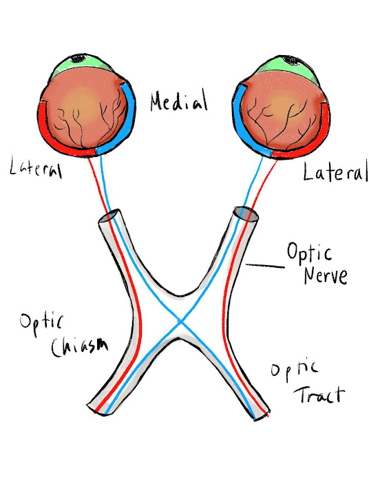
Once inside the middle cranial fossa, the two optic nerves from each eye meet and merge at a structure called the optic chiasm. This crossing point sits right above a part of the sphenoid bone called the sella turcica. At the optic chiasm, the nerve fibers from the inner (medial) part of each retina cross over to the opposite side of the brain, while the fibers from the outer (lateral) part stay on the same side.

This arrangement means that information from the left side of what you see ends up traveling to the right side of your brain, and information from the right side goes to the left side.

After crossing (or not crossing) at the optic chiasm, these nerve pathways are called optic tracts. Each optic tract carries the visual information to a relay center deep in the brain called the lateral geniculate nucleus, or LGN, which is part of the thalamus. From the LGN, the signal is passed to the next set of neurons that send it along special pathways called optic radiations.

The lower parts of what we see are sent along the upper optic radiations, while the upper parts of our view travel along the lower optic radiations. Both paths lead to the primary visual cortex in the occipital lobe at the back of the brain—where the brain finally pieces everything together and creates the clear, detailed images we see every day.


Friday, February 21, 2025

The Eye

Our eyes are one of the most remarkable parts of our body. They help us take in the world around us by detecting light and sending information to the brain through the optic nerve. The brain then pieces these signals together to create the images we see every day.

The bones that form the eye socket, or orbit, are quite intricate. They include seven different bones: the maxilla, zygomatic bone, frontal bone, ethmoid bone, lacrimal bone, sphenoid bone, and palatine bone.

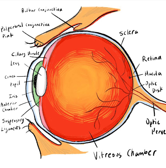
Our eyelids, which are made of soft tissue, act as protective covers for the eye. The white part we see is called the sclera, and it’s lined by a thin, clear layer called the conjunctiva. The cornea is the clear outer layer at the front of the eye. It stays smooth and moist thanks to tears produced by the lacrimal gland. Right behind the cornea is a small space called the anterior chamber, which is filled with a fluid known as aqueous humor.

The colored part of the eye is called the iris, and it determines whether someone’s eyes look brown, blue, green, or hazel. Inside the iris are muscles that control the size of the pupil—the dark circle at the center. The pupil adjusts its size automatically to let in the right amount of light. When it’s bright, the pupil gets smaller so we aren’t overwhelmed by light. In a dark room, it widens to help us see better. These changes happen because of tiny muscles called the sphincter pupillae and the dilator pupillae, which work without us having to think about them.

When light enters the eye, it first passes through the cornea and the aqueous humor. Then, it goes through the lens, which focuses the light, and into a gel-like substance called the vitreous humor. This jelly helps the eye keep its round shape. After passing through all these layers, light finally reaches the retina, which is a special layer full of cells that react to light and turn it into signals for the brain.

In the retina, there are two main kinds of light-sensitive cells: rods and cones. Rod cells help us see in low light and are mostly found around the edges of the retina, giving us peripheral vision. Cone cells, which are concentrated in the center area called the macula, let us see colors like red, green, and blue. There are about 120 million rods and 6 million cones in each eye!

These photoreceptor cells turn light into tiny electrical messages. These messages travel along the optic nerve to the brain. There is one spot on the retina, called the optic disc, where the optic nerve leaves the eye—this spot doesn’t have any light-detecting cells, so it creates a small blind spot in our vision.

Here’s a simple way to find your own blind spot:

  1. Close your left eye.

  2. Hold your left arm straight out and point your left thumb up.

  3. Hold your right thumb up next to your left thumb.

  4. Focus your right eye on your left thumb.

  5. Keeping your gaze on your left thumb, slowly move your right thumb to the right. When your thumbs are about six inches apart, your right thumb will disappear. You’ve just found your blind spot!

Saturday, January 25, 2025

Structure of the Face

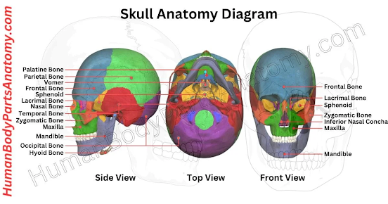
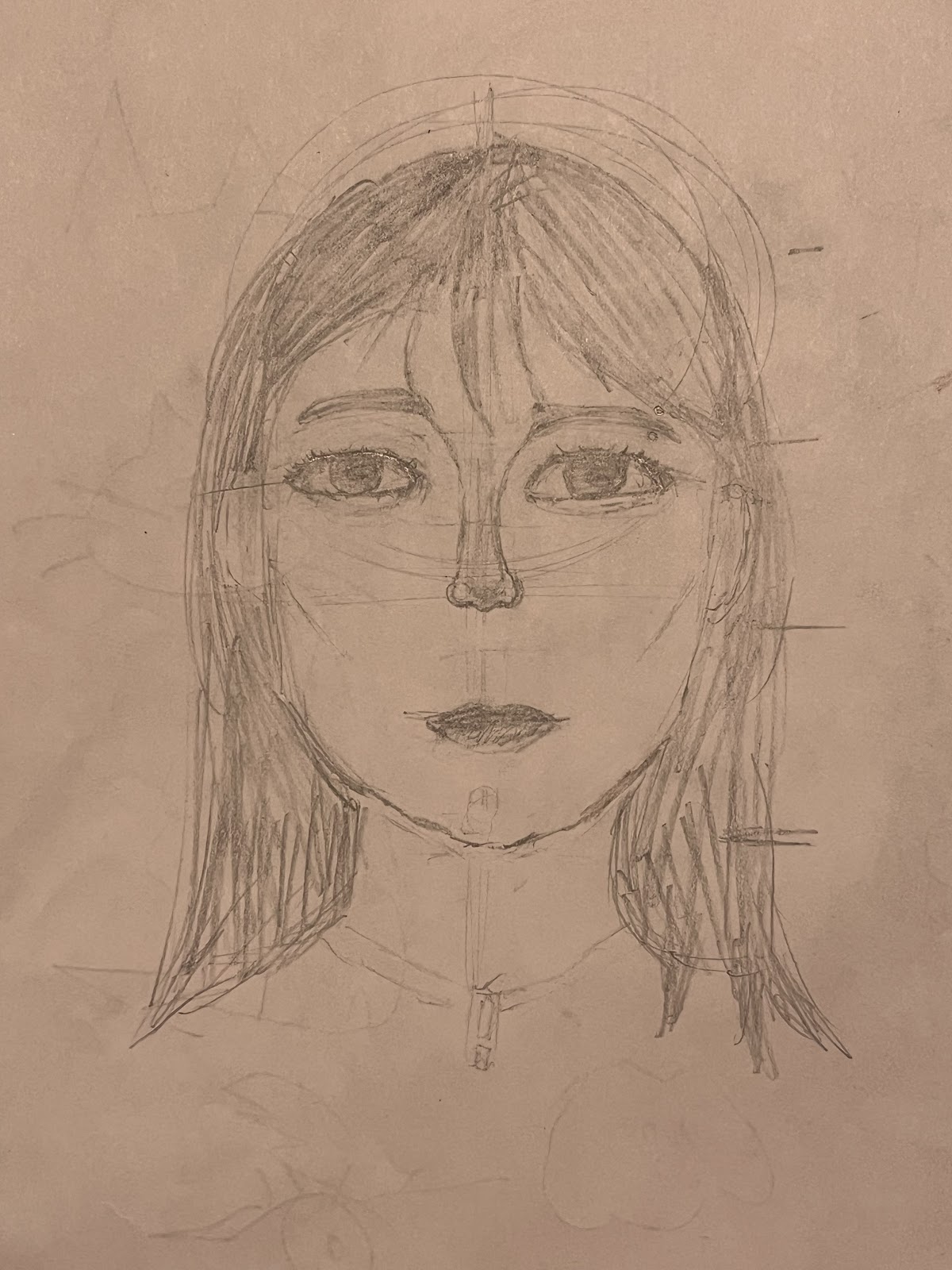
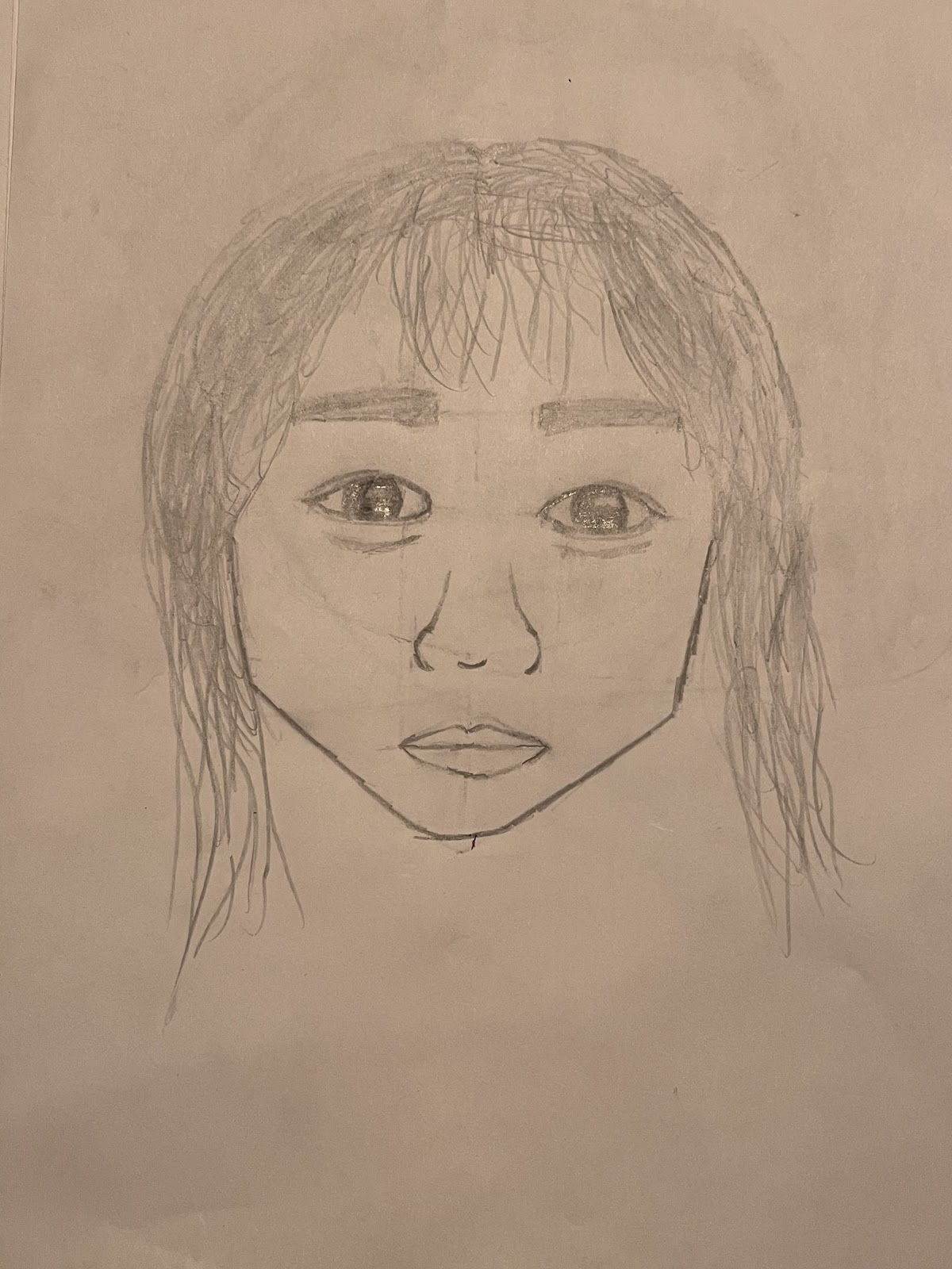
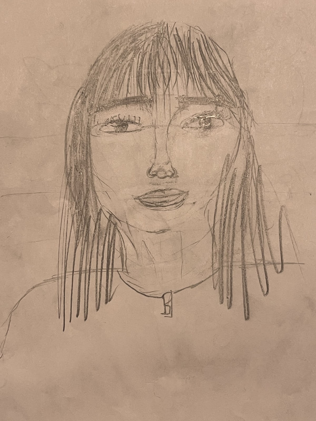
Biology and art are deeply intertwined as biology is the building blocks of art. Common art subjects such as human faces are difficult to capture unless the artist understands the basic structural form beneath the skin. Thus, many artists study anatomy and human physiology. As an artist, I understand the importance of knowing these forms. So, when I volunteered at Sully Community Center to teach a lesson on drawing portraits, I began the session with an overview of skull anatomy. 


I brought a mold of a human skull to help the students visualize the basic forms that make up a face. We studied the skull bones and sutures. I pointed out anatomical features such as the orbit and the mandible to illustrate how small variations in the bones can result in dramatically different faces. Even smaller bones like the nasal bone can impact a person’s facial profile immensely. The kids then brainstormed about how changing a certain bone would impact someone’s face. For example, a wider mandible will result in a wider jaw. This exercise helped them build a solid foundation in determining the bone structure for our reference. 

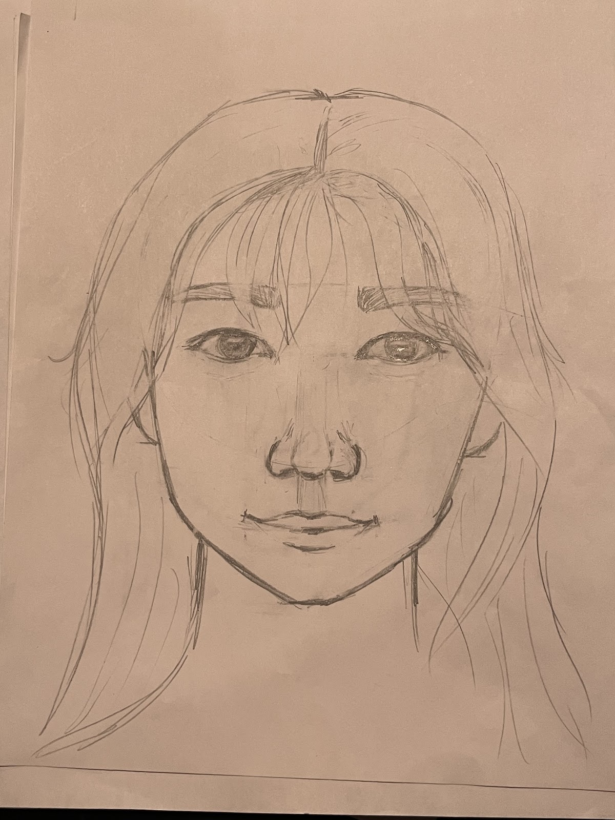
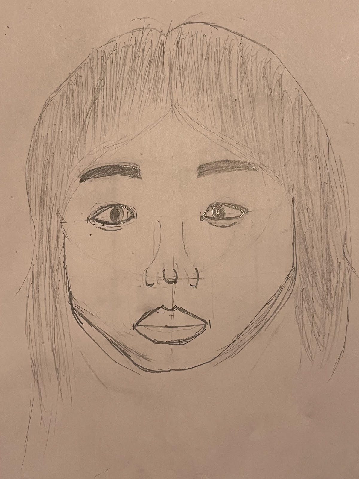
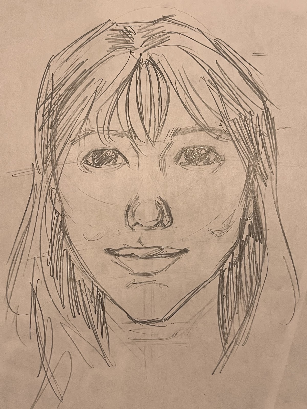
After discussing how variations in these basic bone structures can affect how an artist has to draw the person, we started our drawings. I selected a character from a popular TV show and challenged each kid to try to capture her most accurately. Everyone had tons of fun analyzing her face and trying to determine her facial form and structure. In the end, each kid learned about how different subjects like biology and art can interact with each other and built a solid foundation for the study of the human face/skull.

Monday, December 30, 2024

Nature's Inspiration

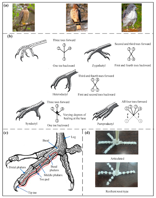
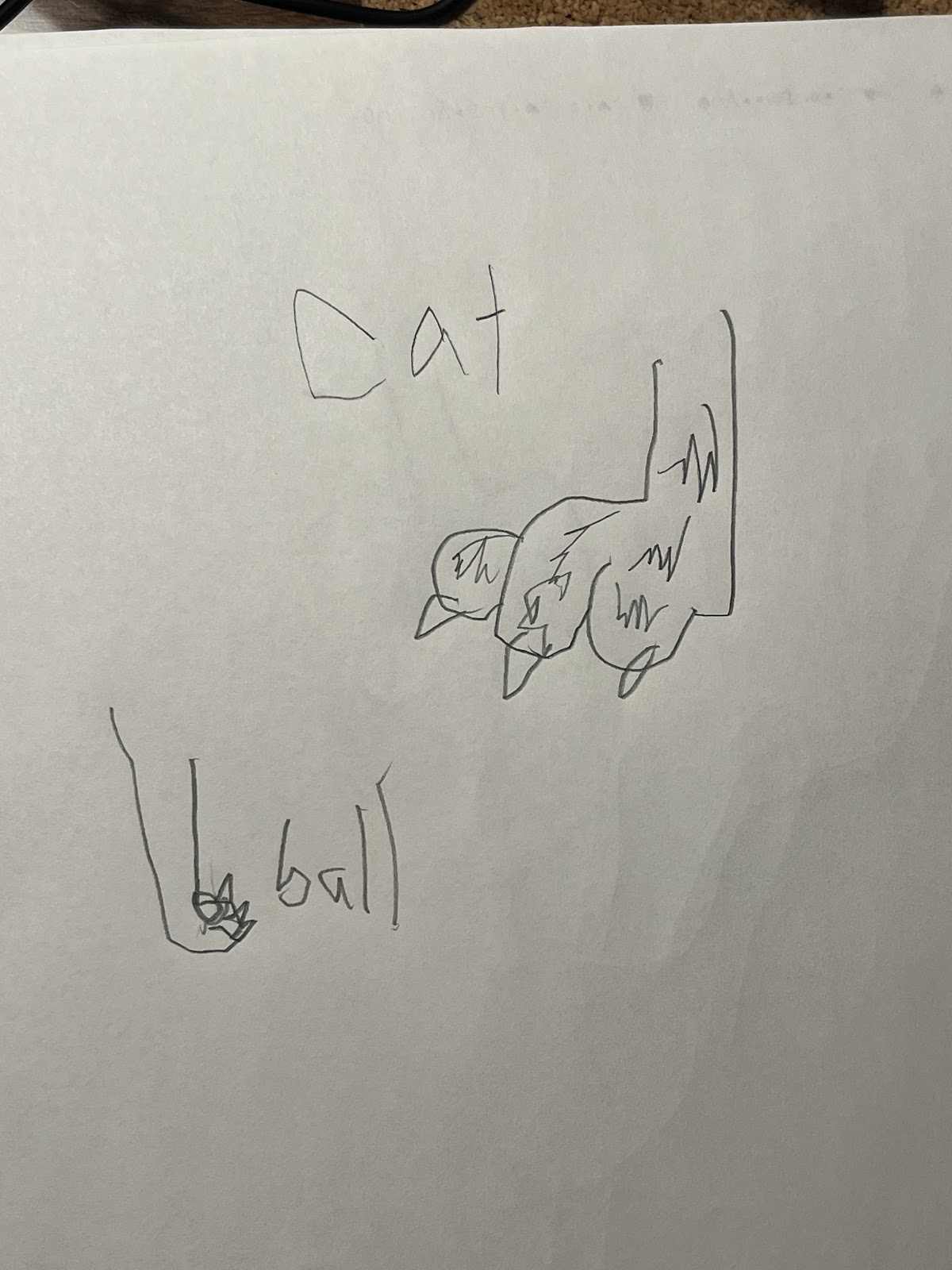
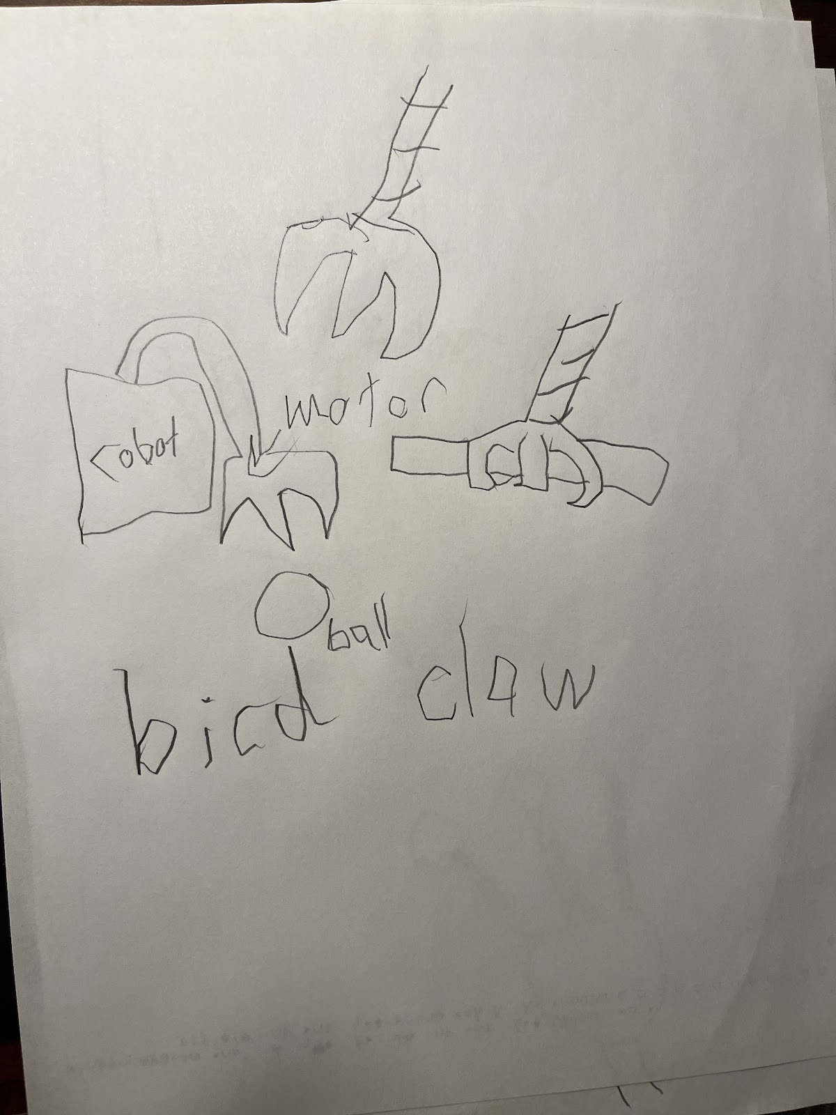
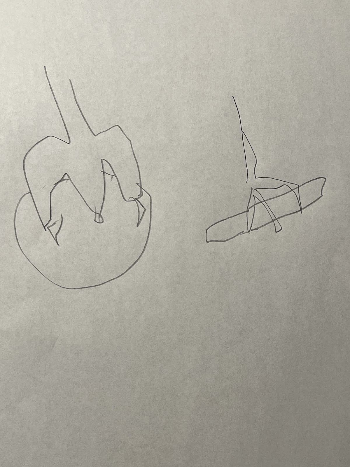
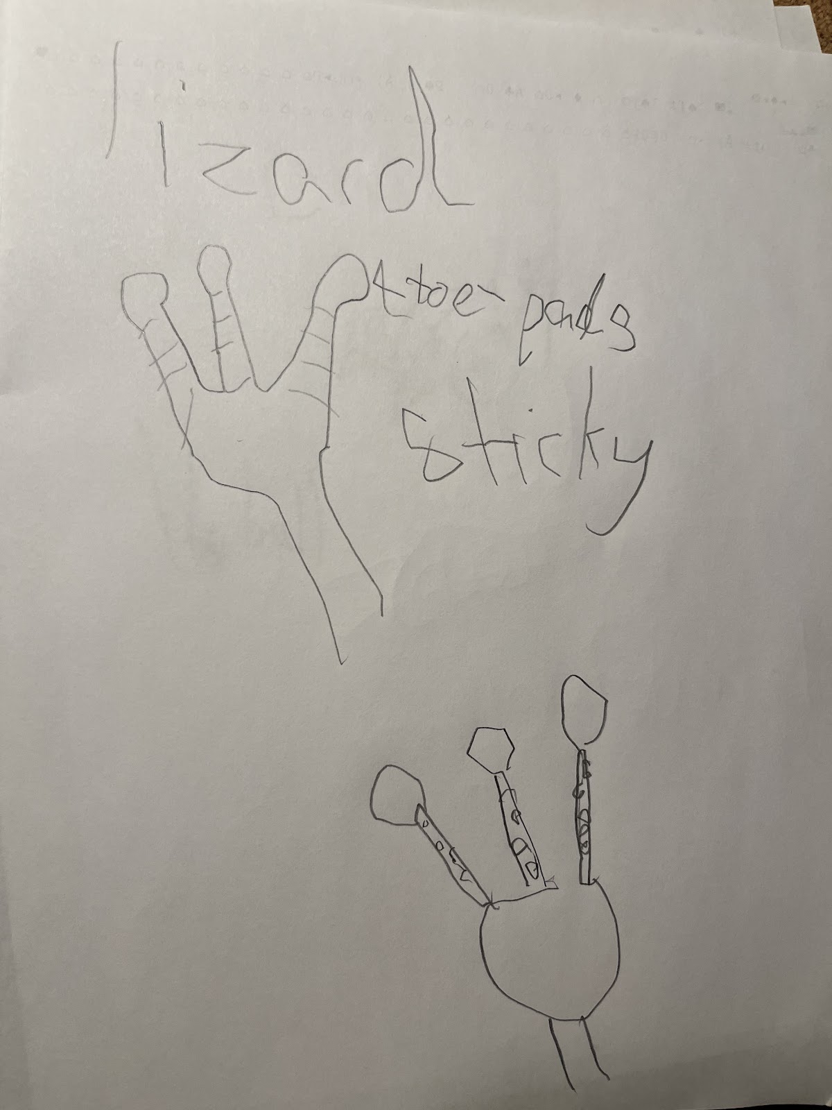
    Over the past weekend, I volunteered at Herndon Fortnightly Library again. This time, we focused on getting inspiration for designs based on animals in real life. Lots of scientists and researchers are able to invent new theories and inventions by observing nature. For example, the first airplane was modeled after a bird! The most important concept isn’t to simply observe the animal anatomy and reproduce it, but to understand why the anatomic feature works and use it to create an original design. I brought pictures of different animals' claws and paws since the final task was to be able to grab a plush ball. The kids could choose from a variety of lizard, cat and bird paws/claws to draw and try to figure out how it worked. After they got a good sense of the mechanics, they had to figure out how to implement their design practically. The main focus of this section was exercising the kids’ creativity and to try to get them to come up with their own original design. Overall, everybody had tons of fun drawing and building from scratch! 

This is an example of how to break down a bird’s claw and figure out its mechanics. This diagram goes in detail with many different types of bird claws such as heterodactyl (2 talons in front, 2 talons in the back), syndactyly (3 talons in front, 1 talon in the back) and pamprodactyl (4 talons in front) claws. 

Sunday, December 8, 2024

Early evolution of small body size in Homo floresiensis

By: Yousuke Kaifu, Iwan Kurniawan, Soichiro Mizushima, Junmei Sawada, Michael Lague, Ruly Setiawan, Indra Sutisna, Unggul P. Wibowo, Gen Suwa, Reiko T. Kono, Tomohiko Sasaki, Adam Brumm & Gerrit D. van den Bergh


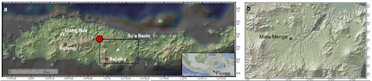
This paper talks about hominid bones unearthed in the So’a Basin in Central Flores, Indonesia. Fragments of a mandible and 6 different teeth belonging to a small hominin were discovered in the sandstone layer of the Mata Menge site. These fossils are dated to between 0.65 and 0.773 Ma. They share morphological similarities to H. floresiensis from Liang Bua (Western Flores) and H. erectus from Java. The fossils seem to be the ancestor of H. floresiensis and are theorized to be dwarf descendents of H. erectus.

Evidence of this link is that the mandible and teeth found at the Mata Menge site are smaller than those found in H. floresiensis from Liang Bua. This hints that these bones may have existed more than 600,000 years before the oldest H. floresiensis fossils found. Unfortunately, due to a lack of cranial bones found in the Mata Menge, there is limited data on body size evolution on Flores. This paper analyzes an adult humerus found later in the same Mata Menge site and concludes that it is morphologically more similar to a Homo species such as H. naledi rather than an Australopithecus. A molar crown also shows similarities to H. erectus rather than early African Homo. These analyses support the fossils being classified as early H. floresiensis. It also provides evidence that between -1.0 and 0.7 Ma, the ancestor of H. floresiensis went through a drastic size reduction from its larger bodied ancestor H. erectus.

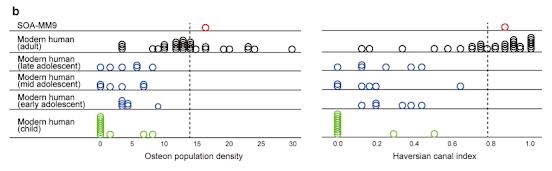
More specific data illustrates the smaller body size of H. floresiensis through the discovered humerus and teeth. Scientists believe the humerus from specimen SOA-MM9 belongs to an adult due to its cortical bone histomorphology. Cortical samples were taken from the mid-posterior shafts of the humerus and compared to a modern human sample. The Osteon Population Density (OPD) and Haversian Canal Index (HCI) of the fossil (OPD = 16.5, HCI = 0.85) were greater than any modern human subadult humerus (OPD = 0.0-8.9, HCI = 0.0-0.63). This indicates that the bone specimen came from a mature adult, not a child. Conditions like osteogenesis imperfecta, which results in a shorter stature, could be ruled out since those with the condition display lower than normal OPD values. The humeral size of the Mata Menge fossil is smaller than any other adult individual of small-bodied fossil hominins such as H. naledi. The Mata Menge humerus’s minimum circumference is 46 mm which is less than the smallest humeri for prehistoric humans (46.5 mm). Its centroid size is also the smallest compared to any known adult specimen of Australopithecus, Paranthropus, H. naledi and H. floresiensis

In the end, researchers concluded that there were at least 4 individual’s remains found in the Mata Menge site, 1 adult, 1 adolescent/ young adult, and 2 children. All 4 of these specimens displayed an extremely small body size for their age. This shows that small body size was not an individual characteristic, but rather a population characteristic. Possible theories  to explain the small stature could be due to a lack of predators. Flores was so isolated that there were no natural predators for early H. floresiensis. Having a larger body size would be a disadvantage evolution wise as larger bodies require more energy and food to sustain itself.


Link to the paper: https://doi.org/10.1038/s41467-024-50649-7 

Creative Common licence: http://creativecommons.org/licenses/by/4.0/

The Peabody Museum

During my time at Yale, I visited The Human Footprint exhibit at the Yale Peabody Museum. The exhibit focuses on how humans and their enviro...