Friday, May 23, 2025

From Fish to Speech

        Hearing is one of the most important senses humans have. We use it to communicate and understand the world around us through sound. The modern human ear developed from a long evolutionary process that began from a fish.

The earliest version of the ear was not actually used for hearing. In prehistoric fish, it began as an organ that helped them sense movement in water and stay correctly oriented. This organ contained fluid and sensory cells that responded to motion. Over time, some fish developed air sacs that further assisted in detecting underwater vibrations. This was one of the early steps towards hearing.

When animals began moving from water to land, hearing had to change as sound travels differently in air than it does water. Amphibians used vibrations from the ground, while reptiles developed a more advanced middle ear. One important bone, called the columella, helped transmit sound and later became the stapes in human ears.

Mammals made hearing even more advanced as they developed three more middle ear bones: the malleus, incus, and stapes. These bones help transfer sound energy into the inner ear more efficiently. Mammals also developed a coiled cochlea, which made certain frequencies to be processed more precisely. Thus, making hearing useful for locating sound and recognizing patterns.

In humans, hearing became deeply intertwined with communications. The ear allowed early humans to listen, respond to sounds, and eventually develop languages. Hearing is part of a larger system involving speech, emotion and social connection. This is the reason why hearing is so important when learning new languages.

The most interesting part of our ears is how they still carry traces of its evolutionary past. Structures that once helped fish sense movement and survive underwater were slowly reshaped into the organs humans now use to hear speech and emotion. The ear demonstrates how evolution often transforms old parts for new purposes. From detecting vibrations in water to understanding language, the human ear is a reminder that our basic senses have deep evolutionary history.

Thursday, May 22, 2025

We are all EARS….

Our ears enable us to hear sound and maintain balance.  They are located over the temporal lobe.  The ear lobes are easily visible, but that’s only a small part of this important organ which is essential for hearing and balance.  There are three main parts to our ear: outer ear, middle ear, and inner ear.  The eardrum (tympanic membrane) separates the outer and middle ear.  

The outer ear is easily visible on the side of the head.  The earlobe is called the pinna.  The external ear canal connects the outside world to the tympanic membrane.  The outer part of the external auditory canal is made of cartilage while the inner two-thirds is made of bone.  Its main function is to transmit sound waves from the surrounding environment to the tympanic membrane.  The fine hairs lining the canal secrets earwax and prevents foreign objects from entering.

The tympanic membrane separates the outer ear from the middle ear and vibrates when sound waves enter the ear canal.  The membrane is divided into a flaccid part (pars flaccida) and a tense part (pars tensa).  It also has a lateral side and a medial side.  The lateral side faces the outer ear while the medial side faces the middle ear.  Part of the facial nerve (CN VII) passes through the membrane. 

The middle ear is located in the temporal bone and consists of three tiny bones called ossicles: malleus (hammer), incus (anvil), and stapes (stirrup).  The handle of the malleus is attached to the tympanic membrane while its head is attached to the body of the incus. The incus is also attached to the head of the stapes.  Finally, the footplate of the stairs is in contact with the oval window.  These ossicles transfer sound vibration from the tympanic membrane to the inner ear.  

In addition, the eustachian tube connects the middle ear with the nasopharynx and helps to equalize air pressure on both sides of the tympanic membrane. The tube is closed at rest but opens with swallowing or yawning so pressure differences can equalize. Thus, when we fly or drive up into the mountains, chewing gum can help to equalize pressure in the middle ear and outer ear.

The inner ear has an outer bony labyrinth and an inner membranous labyrinth. The vestibule is a central bony cavity that connects with the cochlea anteriorly and semicircular canals posteriorly.   

Cochlear:  The cochlear transforms sound waves into electric signals for the vestibulocochlear nerve.  Stapes, one of the middle ear bones, vibrates when sound is transmitted.  The stapes is in contact with the oval window and transmits vibration of sound waves to the cochlear fluid.  The hair cells in the cochlear duct detect the sound vibration and convert it into an electric signal.  The cochlear duct (scala media) is filled with endolymph.  There are also two chambers called scala vestibuli and scala tympani filled with perilymph.  The cochlear duct is positioned between the scala vestibuli and scala tympani.  

Vestibular system (vestibule and semicircular canals):  This system is responsible for balance. There are 3 fluid filled semicircular canals oriented at 90 degrees from each other and help to control balance.  The canals are lined by hair cells which can sense body movement and position.  There are three semicircular canals: anterior, posterior, and lateral.  When the head moves, the fluid and hairs also move.  This movement is converted to an electric signal along the vestibular nerve.  The brain processes this information and then sends signals to muscles to help the body maintain balance.


Friday, April 25, 2025

Variations in the Maxillary Sinus

This paper explores variation in maxillary sinus (MS) anatomy between individuals of East Asian, European, and African ancestry, and how these differences may contribute to health disparities in chronic sinusitis. The MS drains mucus through a small opening called the maxillary sinus ostium (MSO), which is located high on the sinus wall. This positioning makes drainage heavily reliant on mucociliary action, rather than gravity. If mucus can’t drain efficiently, it builds up and can lead to infection. The study used 3D CT scans of 167 adult skulls to measure MS and nasal fontanelle (NF) shape and size, especially the vertical distance from the sinus floor to the MSO.

The results showed that East Asian individuals tend to have significantly taller and larger MSs, with the MS floor positioned farther below the MSO. This increased vertical distance means that mucus has to travel farther to be cleared, possibly increasing the risk of chronic sinusitis. In contrast, individuals of European and African ancestry had shorter MSs and shorter distances between the MS floor and MSO, which may allow for more efficient drainage. Despite these differences, the NF—which is used as a bony landmark for the MSO—was similarly sized and positioned across all ancestry groups, suggesting that surgical approaches to sinus treatment can be standardized.

Importantly, the study emphasizes that these anatomical features do not directly cause disease based on ancestry. Rather, individuals with taller MSs—regardless of ancestry—may be more susceptible to sinus infections. The findings help explain why nonallergic (noneosinophilic) sinusitis is more common in East Asian populations and suggest that internal anatomy, along with socioeconomic and environmental factors, contributes to global health disparities.

The authors conclude that anatomical variation, especially MS height, may play a role in sinusitis risk and should be considered in both clinical treatment and future research. This paper highlights the need to study not just pathology or healthcare access, but also how normal anatomical differences between populations might shape disease vulnerability.

This is the paper I read:

https://anatomypubs.onlinelibrary.wiley.com/doi/full/10.1002/ar.24644 

Wednesday, April 9, 2025

The Anatomy of the Sinuses

 The paranasal sinuses are four paired, air-filled cavities in the face, arranged around the nasal cavity. All of them drain into the nasal cavity. The four sets of paired sinuses are the maxillary, frontal, sphenoid, and ethmoid sinuses. The drawing below displays them:

The paranasal sinuses develop in the fetus as small outpouchings from the nasal cavity that grow into the surrounding bones. After birth, they continue to develop until early adulthood. The sphenoid sinuses are unique to humans and primates. The maxillary sinuses are the largest, with an average volume of 15 mL in adults. The frontal sinuses are the most superior in position. The ethmoid sinuses are made up of multiple small air cells divided into anterior, middle, and posterior groups, and they vary in size.

The exact functions of the paranasal sinuses are not fully understood, but they have several roles. They help fight infection by producing mucus that drains into the nasal cavity and clears germs. They also add moisture to the air we breathe. They help absorb impact and protect the brain in case of head or face trauma. They also increase the resonance of our voices and, because air is lighter than bone and muscle, they make the head lighter overall.

The maxillary sinuses sit beneath the eyes in the maxillary bones. They are small at birth but grow rapidly between ages 0–3 and again between 6–12 years. In adults, they reach about 15 mL and are the largest sinuses. They are also the most common site of sinusitis. They drain into the nasal cavity through the hiatus semilunaris into the middle meatus. The floor of the maxillary sinuses is formed by the alveolar process of the maxilla and lies close to the roots of the upper teeth, which means that dental infections, such as abscesses, can spread into these sinuses. The osteomeatal complex connects the frontal, anterior ethmoid, and maxillary sinuses to the middle turbinate.

The frontal sinuses are located in the frontal bone above the eyes and have a triangular shape. They are the most superiorly located sinuses. Absent at birth, they begin forming around age two but do not become visible until about ages five or six. Their drainage pathway can vary. Fluid usually flows through the frontal recess, then either into the hiatus semilunaris and middle meatus or directly into the middle meatus. Like the maxillary sinuses, they are also a common site of infection.

The sphenoid sinuses are found behind the eyes and are the deepest sinuses. They occur only in humans and primates. They are absent at birth but begin to appear around age two, maturing between ages twelve and fourteen. They drain through the sphenoethmoidal recess into the nasal cavity. Important structures, such as the internal carotid artery and the optic nerve, lie close to the sphenoid sinuses. Surgeons can pass instruments through the nose into the sphenoid sinus and then into the hypophyseal fossa to treat pituitary adenomas. This approach, called endoscopic trans-sphenoidal surgery, provides a minimally invasive method to remove pituitary tumors.

The ethmoid sinuses are located between the eyes and behind the nose. They appear as small pockets of air cells separated by thin septa. These sinuses are present at birth and mature around age twelve. Their total number varies, ranging from four to seventeen. They are divided into anterior, middle, and posterior groups. The anterior group drains via the hiatus semilunaris into the middle meatus, the middle group drains into the lateral wall of the middle meatus, and the posterior group drains into the lateral wall of the superior meatus.


Tuesday, March 25, 2025

The Life of Paul Farmer

I came across an article in the American Anthropologist Journal about medical anthropologist Paul Farmer. His life story is inspiring and pushes everyone to start thinking about how they can help others. Paul Farmer believed medicine had to start with people’s lives. Born in 1959, he grew up in unusual places like a converted school bus and a houseboat in Florida. He went to Duke University, where a medical anthropology class changed how he viewed medicine. Instead of studying science only in a lab, Farmer wanted to understand how history, politics, and culture shaped people’s health.

In the 1980s, he worked in rural clinics in Haiti, where malnutrition and preventable diseases were part of daily life. Rather than just observing, he built partnerships with local leaders such as Father Fritz Lafontant and the people of Cange. Together they founded Partners In Health (PIH), which became a global network proving that tuberculosis and HIV could be treated among the poor. Farmer called this approach a “preferential option for the poor.” It meant hiring and training local health workers who visited patients at home, brought medicine, and made sure basic needs like food and housing weren’t ignored.

Farmer was never afraid to call out injustice. At Harvard Medical School, he realized there were excess medical supplies in Boston hospitals while his patients in Haiti went without the basics. He called the deaths he witnessed “stupid deaths,” meaning deaths caused by diseases that were preventable and treatable. His books such as Pathologies of Power and Infections and Inequalities explained this as structural violence, showing how poverty and racism quietly but powerfully harm poor and vulnerable people.

Even as PIH expanded to countries like Peru and Rwanda, Farmer stayed close to his patients. In Boston, he treated the homeless, and in Africa, he helped fight the Ebola epidemic when many global health authorities had given up. Farmer believed that solidarity meant showing up everyday. He once said, “You just don’t leave.” That commitment inspired a generation of students and doctors to think differently about health as a human right.

Farmer was also known for his generosity and humility. He often gave away his possessions, even borrowing money just to hand it to someone else in need. Before an important meeting, he once fixed his only suit with a black marker. He cooked meals for family and took time to listen to everyone he met. After his lectures, he would stay for hours to answer questions from students and strangers.

When he died in Rwanda in 2022, Farmer was still doing what he had always loved: visiting patients, mentoring young doctors, and sharing jokes in the hospital wards. His gravestone reads, “Paul Farmer—Was of Use.” For the communities he served, that is more than true.


Tuesday, March 11, 2025

Evolutionary Impact of Social Isolation

One of the greatest mysteries in human evolution is how the Neanderthals went into extinction ~40 thousand years ago. This paper discusses the genome of a Neanderthal called “Thorin,” found in Grotte Mandrin in Mediterranean France. Thorin’s remains include a mandible and 31 teeth from between 52,900 and 48,050 years ago. These remains are linked to the PNII lithic industry, one of the final Neanderthal cultural phases in the region. Genomic analysis shows that Thorin belonged to a previously unknown Neanderthal lineage that diverged from other late European Neanderthals around 100,000 years ago and then remained genetically isolated for about 50,000 years. This isolation is unusual because other late Neanderthals were thought to be part of a single connected population.

Genetic analysis supports the isolation of this Neanderthal lineage. Thorin’s genome showed no signs of gene flow with other Neanderthals, including those living nearby. His mitochondrial DNA was most closely related to individuals from Gibraltar and Poland, and his Y chromosome also diverged early. Thorin also had high levels of homozygosity, with around 7% of his genome in long homozygous stretches, indicating recent inbreeding and a small group size. These findings suggest that his population had limited social contact with other Neanderthals. Interestingly, this isolated group also had a distinctive cultural identity, with stone tools that differed from the better-known Mousterian and Châtelperronian traditions in nearby areas.

The researchers conclude that Thorin's population likely represents one of the last Neanderthal groups in Europe, living in isolation until around 42,000 years ago. Their genetic and cultural separation may have made them more vulnerable to extinction. Unlike modern humans, who built wide social networks and exchanged genes across regions, these Neanderthal groups were fragmented and possibly avoided interbreeding—even with other Neanderthals. This paper adds more knowledge to the debate about Neanderthal extinction, suggesting that social and genetic isolation may have been just as important as external factors like climate or modern human expansion.

This is the paper I read: https://www.cell.com/cell-genomics/fulltext/S2666-979X(24)00177-0


Friday, February 28, 2025

The Optic Pathway

The optic nerve, which is known as the second cranial nerve, plays an essential role in helping us see. It carries the information collected by the retina and delivers it to the primary visual cortex in the brain, where our brain makes sense of what we are looking at.

Outside the brain (extracranial):
Inside the retina, special light-sensitive cells called photoreceptors turn light into tiny electrical signals. These signals are passed on to cells called bipolar cells, which release a chemical messenger called glutamate. This chemical activates the retinal ganglion cells, which create action potentials—tiny electrical impulses that are the language of our nervous system.

The long extensions (axons) of the retinal ganglion cells come together to form the optic nerve. This nerve leaves the protective bony eye socket through a small passage called the optic canal and then enters the skull’s middle cranial fossa, a space near the pituitary gland.

Inside the brain (intracranial):
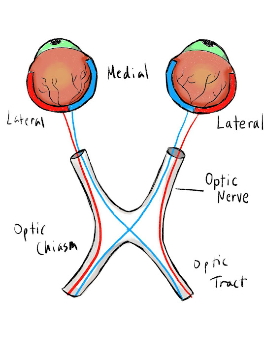
Once inside the middle cranial fossa, the two optic nerves from each eye meet and merge at a structure called the optic chiasm. This crossing point sits right above a part of the sphenoid bone called the sella turcica. At the optic chiasm, the nerve fibers from the inner (medial) part of each retina cross over to the opposite side of the brain, while the fibers from the outer (lateral) part stay on the same side.

This arrangement means that information from the left side of what you see ends up traveling to the right side of your brain, and information from the right side goes to the left side.

After crossing (or not crossing) at the optic chiasm, these nerve pathways are called optic tracts. Each optic tract carries the visual information to a relay center deep in the brain called the lateral geniculate nucleus, or LGN, which is part of the thalamus. From the LGN, the signal is passed to the next set of neurons that send it along special pathways called optic radiations.

The lower parts of what we see are sent along the upper optic radiations, while the upper parts of our view travel along the lower optic radiations. Both paths lead to the primary visual cortex in the occipital lobe at the back of the brain—where the brain finally pieces everything together and creates the clear, detailed images we see every day.


Makeup and Face Transplants

A fascinating question Fay Bound-Alberti asks in The Face: A Cultural History is about face transplants and how much of your identity is ti...